
Cancer researchers have to deal with some of nature’s ugliest diseases, but they do find bits of beauty along the way – and that beauty is the focus of an art walk presented by Seattle’s Fred Hutchinson Cancer Research Center on Thursday.
The event features scientific images that were captured by researchers at Fred Hutch, and will be put on display from 5:30 to 8 p.m. in the Mundie Courtyard on the research center’s South Lake Union Campus, at 1100 Fairview Ave. N.
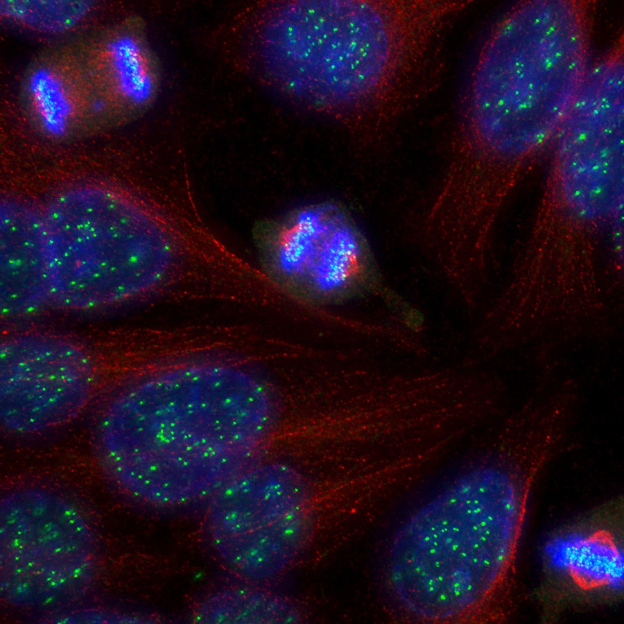
One picture focuses in on a single dividing tumor cell from a human brain, glowing red with bright blue spots called kinetochores. Another shows a burst of brain cells in the cerebral cortex of a developing mouse, illuminated in blue, green and fuchsia.

Cecilia Moens, a developmental scientist at Fred Hutch, emphasized that the pictures weren’t taken because they’re pretty.
“The loveliness is irrelevant to the science,” she told GeekWire. “It’s unimportant that they be appealing, but sometimes they are.”
The selected images highlight the aesthetic side of photomicrography, but they also shed light on the studies being conducted at the Hutch. For example, consider this starburst image from Mitsutoshi Nakamura, who works in the Hutch’s Parkhurst Lab.

“That’s a laser-induced wound in an early fly embryo,” Moens said. The patterns of actin filaments that form around the wound help Nakamura and his colleagues figure out the process by which cells heal themselves.
Actin also shows up in a squiggly image of a cell’s cytoskeleton, also from the Parkhurst Lab. The photomicrograph provided the first evidence of a protein (shown in yellow) that can link up actin filaments (green) link up with microtubules (red) to coordinate their roles in forming cellular scaffolding.
Understanding how such cytoskeletons are formed can help scientists tease apart the tangled mysteries behind the growth and spread of cancer cells.

Thursday’s art walk will feature a variety of two-dimensional and three-dimensional works, and researchers from the Hutch will talk about the works and the science behind them.
Although scientists may not consider themselves artists, Moen acknowledged the parallels between the two types of endeavors.
“Art and science have the same ultimate goal, which is to understand the world,” she said.
More than 500 people have RSVP’d for Thursday’s event, but it’s not too late to add your name to the list.


